

TOPIC 4: COORDINATION ~ BIOLOGY FORM 5
TOPIC 4: COORDINATION ~ BIOLOGY FORM 5
COORDINATION
Coordination is a linking together of the functions of different organs so that they work at a fine time and rate required by the body.
Coordination is achieved through a nervous and endocrine or hormonal system.
DIFFERENCE BETWEEN NERVOUS AND ENDOCRINE
THE NERVOUS SYSTEM
Nervous system involves five main components: These are
1.Stimulus: A change in the external or internal environment e.g. touch, pain, smell and sound.
2.Receptor: A structure which detects the change in the environment e.g. eyes, ears, nose, skin and tongue (sense organs).
3.Coordinator: An organ which receives message from receptor and use the message to coordinate the activities in the body e.g. brain, spinal cord, and messages received (impulses).
4.Effectors: An organ which is controlled by the brain or spinal cord to bring about appropriate response e.g. muscles and glands.
5. Responses: A body activity provoked by a stimulus e.g. pulling away a hand when accidentally one touches a hot object.
Nervous system is made up of interconnected nerve cells or neurones.
Properties of neurones which distinguish it from other cells.
Excitability– highly capable of responding to stimuli.
Conductivity- capable of conducting massage along it.
Functions of the nervous system.
Receive stimuli.
Convert stimuli into the form of electrical impulses.
Transmit the impulses over a considerable distance.
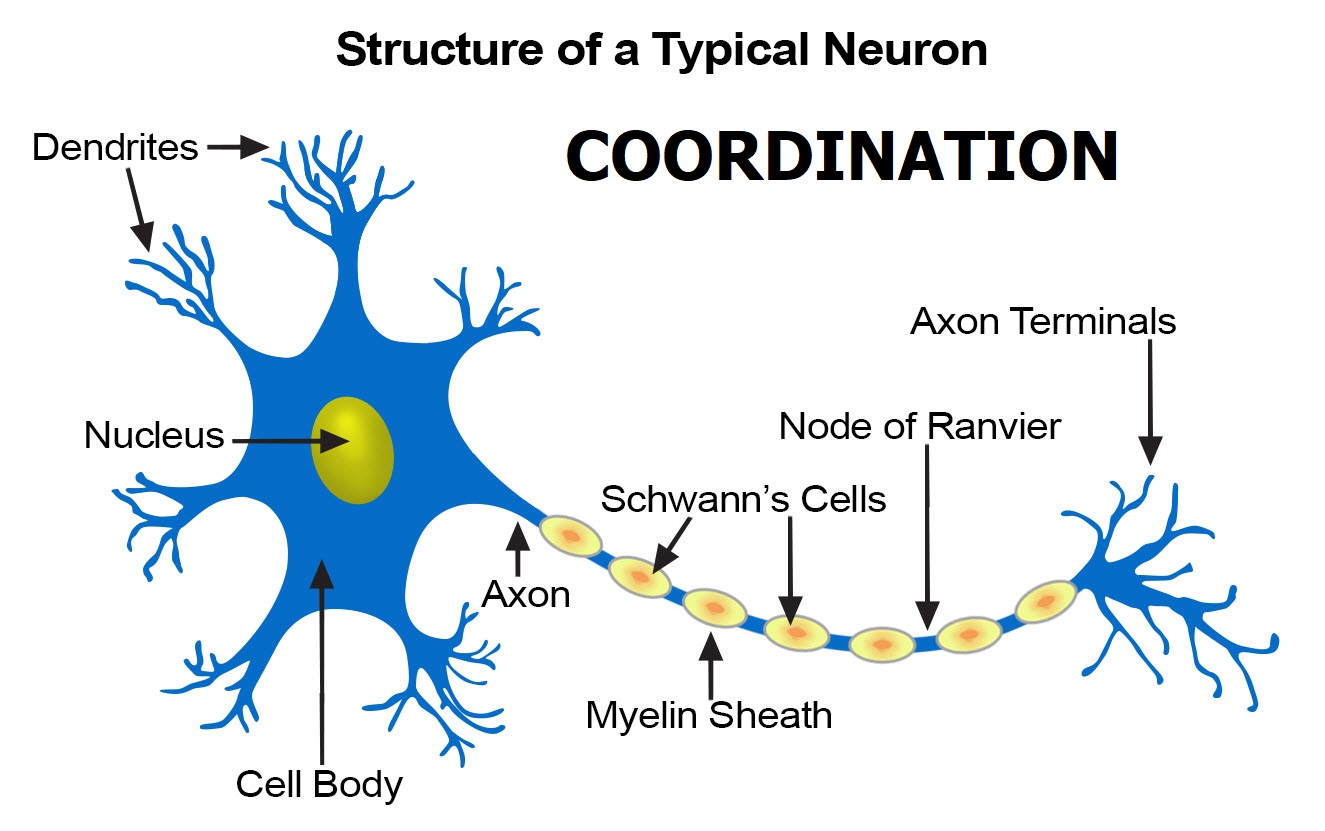
STRUCTURE OF NEURONES.

Neurones are the basic units of nervous system.
1. Cell body
Contains the nucleus, mitochondria and other organelles along with Nissl’s granules.
The prominent groups of ribosomes for protein synthesis.
The cell body has slender finger – like processes called dendrites which connect with neighboring nerve cells.
2. Nerve fibre
In nerve fibre we have axon and dendrites.
Axon transmits impulses away from the cell body and the dendrites transmit impulses towards cell body.
3. Schwann cell.
Some vertebrates are associated with this cell.
The Schwann cell membrane is wrapped repeatedly around the neurone forming a fatty layer known as myelin sheath.
This is important for two reasons.
The myelin sheath is surrounded by a thin layer known as neurilemma which is not part of the neurone but the membrane of another cell (Schwann cell) or glial cells. Each axon is filled with axoplasm.
Nerve cells are referred as unipolar, bipolar, multipolar, etc. according to how many dendrones project from the cell body.
Intermediate nerve cells are bipolar has two unconnected fibres a Dendron and axon which enter and leave at opposite sides of the cell body.
Nerve fibres are bonded together to form nerves some carry only sensory fibre and are known as sensory nerves while other carry a mixture of motor and sensory fibres and are called mixed nerve.
CLASSIFICATION OF NERVES
AFFERENT OR SENSORY NEURONE
Transmit impulse towards the brain or towards the spinal cord.
The dendrite terminates into sense organ.
The cell bodies of sensory cells are found to one side of the main nerve fibre and are frequently collected together in a ganglion.
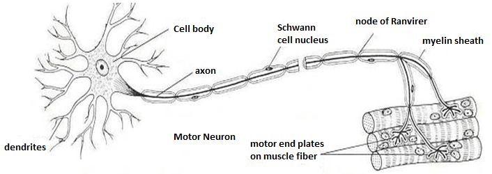
EFFERENT OR ASSOCIATION NEURONE (MOTOR)
Carries impulses from the brain or from the spinal cord to the effectors organ.
It is situated in the brain or spinal cord.
The terminal part of axon is situated in an effectors organ.
Infantile paralysis or poliomyestitis (polio) sometimes cause destruction of efferent neurons leading to tissue atrophy of skeletal muscles.
When this happens no nerve impulse will be reaching the muscles and as such relaxation will no longer be possible.
This condition is called paralysis.
Under such a condition the muscles diminished in size due to decrease, should paralysis vital organs, the animal dies.
1. RELAY OR INTERMEDIATE NEURONES.
Link afferent and efferent neuron.
The terminal part of the dendron receives the impulses from the terminal part of the axon or relay neuron are passed into the dendrites of a motor neuron.
Relay neuron eternal entirely use in the spinal cord and in the brain.

SPEED OF TRANSMISSION IN NEURONES.
The speeds at which the message can be carried depend largely on two things.
In general the larger the diameter the more rapid impulses travel along it.
Myelinated neurone can carry impulses much faster than a (non myelinated neurone.)
The effect of the myelin sheath is to speed up the transmission of a nerve impulse without a need for giant axons.
NERVE IMPULSES.
The nerve impulse is a minute electrical event which is the result of charge differences across the membrane of the nerve fibre.
THE RESTING NEURONE
The membrane of the axon like any other cell surface membrane is partially permeable.
It is the difference in permeability of this membrane to sodium and potassium ions which sets neurones apart from other cells and gives them their special conducting properties.
The axon membrane is relatively impermeable to sodium ions but quite freely permeable to potassium ions.
It also contains a very active sodium/ potassium pump which use ATP to move sodium/potassium pump which uses ATP to move sodium ions out of the axon and potassium ions in.
The function of this is to reduce the concentration of sodium ions inside the axon. They are pumped out and cannot diffuse back in.
At the same time potassium ions are moved in but then diffuse out again along the concentration gradient.
As a result the inside of the cell is left slightly negative charged relative to the inside.
It is polarized. There is a potential difference across the membrane of – 70mv which is known as the resting potential.

NB: The resting potential of the axon is maintained by the sodium pump the relative permeability of the membrane and the movement of potassium ions.
THE ACTIVE NEURONE
When the impulse travels along an axon there is a change in permeability of the cell surface membrane to sodium ions. This change occurs in response to a stimulus.
When a neuron is stimulated the axon membrane shows a sudden and dramatic increase in its permeability to sodium ions specific sodium channels or sodium gates open up, allowing sodium ions rich in both concentration and electrochemical gradients.
As a result the potential difference across the membrane is briefly released, the cell becoming positive on the inside with respect to the outside.
The depolarization lasts about 1 millisecond.
The potential difference across the membrane at this point is about +40mv.
This is knows as action potential.
At the end of this brief depolarization, the sodium channels close again and the excess sodium ions are rapidly pumped out by the sodium pump.
Also the permeability of the membrane to potassium ions is temporarily increased so that the excess potassium ions diffuse in along an electrochemical gradient.
It takes a few millisecond (about 3 ms) before the resting potential is restored and nerve fibre is ready to carry another impulse.
It is this refractory period which ensures that the nerve impulses only transmits in one direction until the resting potential is restored, the part of the nerve fibre that the impulse has just left and cannot conduct another impulse so the impulse can only continue travelling in the same direction.
REFRACTORY PERIOD.
Refractory period is the period of in excitability that accompanies the recovery phase of the axon.
ACTION POTENTIAL.
The action potential is brought about by the movement of sodium ions through the opened sodium channels.
The resting potential is restored by the closing of channels the action of the sodium pump removing excess sodium ions and the movement of potassium ions along an electrochemical gradient.
THE THRESHOLDS
The threshold for any nerve fibre is the point at which sufficient sodium channels open such that the rush of sodium ions into axon is greater than the outflow of potassium ions.
Once the threshold has been reached the action potential occurs. The size of this action potential is always the same. It is all – or- nothing response.
STUDY QUESTIONS.
a) by using large, well labeled diagram explain the two types of cells from the two different systems of a body of a mammal.
COORDINATION
b) The all –or– nothing law states that “The response of an excitable unit is independent of the intensity of the stimulus”.
Clearly explain
It means that the action potential is either generated in which case it is always the same or it is not generated when the stimulus is too small.
COORDINATION
lower-roman;margin-left: -36.75pt;text-decoration: none;vertical-align: baseline”>
lower-roman;margin-left: -36.75pt;text-decoration: none;vertical-align: baseline”>
COORDINATION
lower-roman;margin-left: -36.75pt;text-decoration: none;vertical-align: baseline”>
lower-roman;margin-left: -36.75pt;text-decoration: none;vertical-align: baseline”>
lower-roman;margin-left: -36.75pt;text-decoration: none;vertical-align: baseline”>
Osmotic pressure of axoplasm.
COORDINATION
a) Explain why a nerve is always indirectional
i) spatial summation ii) Temporal summation
b) Explain the usefulness of these concepts as far as nerve impulse transmission is concerned.
Examine the ionic movements’ across the neurons –membrane and the associated phenomena.
COORDINATION
REFRACTORY RERIOD
Refractory period is …..
The refractory period is the time it takes for an area of the axon membrane to recover after an action potential that it takes for the ionic movement to reporalize.
COORDINATION
This is brought about by the sodium pump and membrane permeability to potassium ions.
For the first millisecond or so after the action potential it is impossible to restimulate the nerve fibre the sodium channels are completely blocked and the resting potential not yet been restored.
COORDINATION
This is known as the absolute refractory period.
After this, there is a period at several milliseconds drawing which the nerve fibre may be stimulated, but it will only respond to a much longer stimulus than before.
COORDINATION
The threshold has effectively been raised. This is known as relative refractory period.
IMPORTANCE OF REFRACTORY PERIOD.
The refractory period is important in the functioning of the nervous system as a whole.
It limits the frequency with which impulses may flow along a nerve fiber to 500 – 1000 each second.
COORDINATION
It also ensures that impulses flow only in one direction along nerve making it possible to have motor and sensory system with no internal confusion.
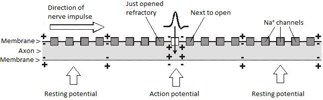
PROPAGATION OF THE NERVE IMPULSE.
So far we have considered the action potential as isolated events in one area of a nerve fibre.
In fact once an action potential has been set up in response to a stimulus; it will travel the entire length of the fibre, which may be many centimeters or even meters long.
COORDINATION
The movement of the nerve impulse along the axons the result of local currents set up by the ion movements at the action potential itself.
They occur both in front and depolarize the membrane sufficiently to the sodium channels to open in front of the action potential as shown below.
(The refractory period prevents the sodium channels opening between the spikes.)
COORDINATION
In this way impulse to be continually propagated in the required direction.
The result of these ions movement is to depolarize the membrane sufficiently to set up a new action potential in on one direction only.
It cannot go backward due to the refractory period.
COORDINATION
COORDINATION

COORDINATION
SALTATORY CONDUCTION.
In myelinated vertebrate nerves, the mechanism of propagation is strongly more complex.
Ions can only pass freely into and out of the axon at the nodes of ranvier which are about 1mm apart.
This means that action potential can only occur at the nodes of ranvier and so they appear to jump from one node to the next as the diagram shows.
COORDINATION
The effects of this is to speed up transmission as the concentration movements associated with the action potential occurs much less frequently taking less time.
This condition is known as saltatory condition from the Latin verb which means to jump.
COORDINATION

LINKING THE SYSTEM
The nerves are basic units of the nervous system adopted for the rapid passage of electrical impulses to inter communicates.
COORDINATION
Receptors must pass their information into the sensory nerves, which in turn must relay the information to the central nervous must be communicated to the effectors organ so that action can be taken.
COORDINATION
Whenever two nerve cells meet, they are linked by synapse as shown in the figure below.
Every cell in the central nervous system is covered with synaptic knob from other cells several hundred some cases.
Neurons never actually touch their target cells so a synapse is a gap between two nerve cells with the nerve message must be somehow crossing.
COORDINATION
The electrical nature of the nerve impulse as detected long before to its could be accurately recorded and measured similarly it was suspected that transmission at the synapses was not electrical but chemical long before the electrons microscope and other technique could demonstrate this clearly.
Once the structure at the synapse had been seen using the electron microscope, the synapse gap would be measured. This settled the argument.
The gap is simply too wide for an impulse or the size of an axon potential to jump across. Synaptic transmission had to be chemical and all the available evidence confirms this.
THE SYNAPSE AT WORK
The arrival of an impulse at the synaptic knob increases the permeability at the synaptic membrane to calcium ions.
COORDINATION
Calcium ions therefore move into the synaptic knob along concentration gradients the effect of these calcium ions is to cause the synaptic vesicles containing transmitters’ substance to move to the pre-synaptic membrane.
Each vesicle fuses with the membrane and release the transmitters.
Some of the vesicles fuse with the membrane and release the transmitters.
COORDINATION
Some of the vesicles fuse with the membrane and release the transmitter substance into the synaptic cleft.
The transmitters diffuse across the gap and become attached to the specific protein receptor sites on the post synaptic membrane.
As a result, ions channels are opened and there is usually a local depolarization and influx of sodium ions, causing an excitatory post synaptic potential (EPSP) to be set up.
COORDINATION
If there are sufficient of the potential the positive charge in the postsynaptic cell build up to the threshold level and an action potential in set up this then travels on a long the post synaptic neuron.
In some cases the transmitters have the opposite effect channels allowing the inward movement of negative ions are opened, in the post synaptic membrane, which makes the inside more negative than the normal resting potential.
An inhibitory post synaptic potential results, which makes it less likely than an action potential occur in the past synaptic fibre.
COORDINATION
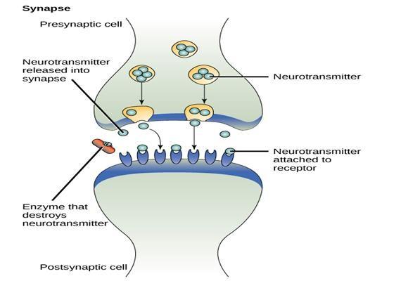
Once the transmitter has its effects it is destroyed by enzymes.
This is very important because unless the transmitter is removed from the synaptic cleft subsequent impulses would have as effect, as the receptors on the post synaptic membrane would the entire bound.
THE TRANSMITTER SUBSTANCES.
The most common transmitter substance found at the majority of synaptic is acetylcholine (Ach).
It is synthesized in the synaptic knob using ATP produced in many mitochondria present.
Nerves using Acetylcholine has done its job it is very rapidly, hydrolyzed by the enzymes cholinesterase.
This ensures that it no longer affects the post synaptic membrane, and it also releases the components to be recycled they pass back into the synaptic knob and are resynthesized into acetylcholine
COORDINATION
Some vertebrates’ nerves particularly those of the sympathetic nervous system produce noradrenaline in their synaptic vesicles and are known as adreneigic nerves.
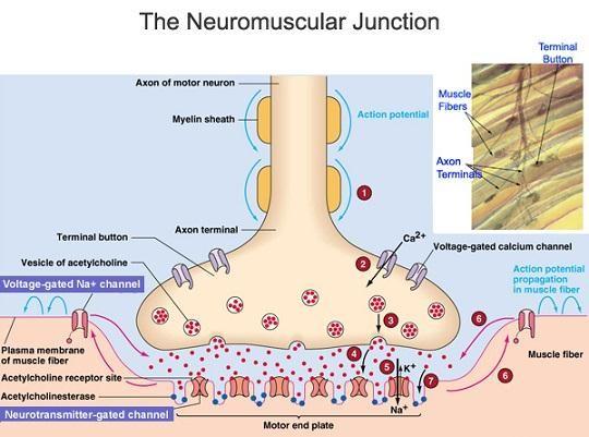
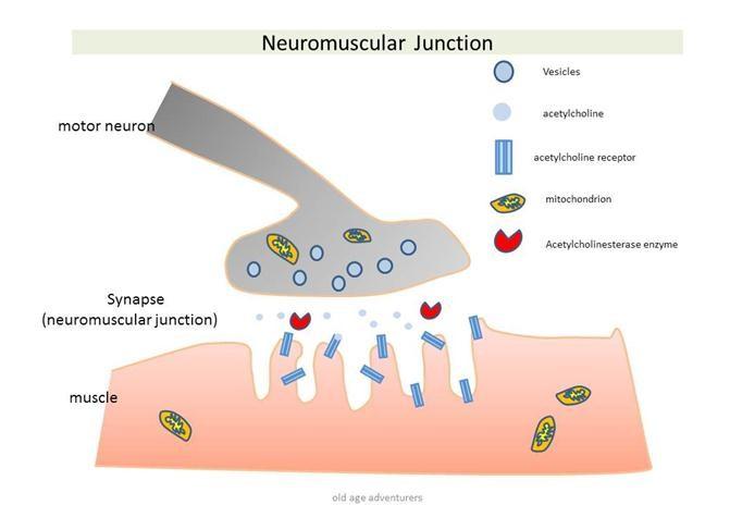
NEUROMUSCULAR JUNCTION
Nerves have to communicate not only with each other but with receptors and effectors as well.
Motor nerves need to communicate with muscles. Where a motor nerve and muscles fibre meet a special kind of synapse is formed known as a neuromuscular junction.
The membrane of the muscle fibre is very folded in this region and forms a structure known as an endplate to which the end of the motor nerve joins.
COORDINATION
Electron microscope shows us that the structure of the neuro muscular junction is remarkably similar to that of any other synapse as the figure below shows.
The end of the motor neurons is full of mitochondria and synaptic vesicles which contain acetylcholine.
It appears that when an impulse arrives at the end of the motor neurone acetylcholine is discharged into the synaptic cleft.
As a result of its effect on the postsynaptic membrane an end potential is set up which can be recorded, If sufficient end plate potentials are set up on action potential is fired off on the muscle fibre spreading through the tubules and leading to a contraction of the muscle.
COORDINATION
COORDINATION

COORDINATION AND CONTROL OF NEURONES SUMMATION AND FACILITATION
Neurons interact in a variety of complex ways.
Sometimes single nerve fibre will carry an action potential to a synapse with another cell and transmission.
But in many cases the situation is much more complex than this.
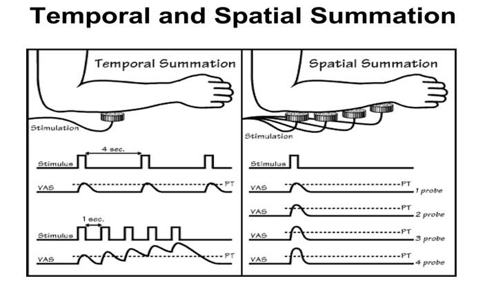
Often a single synaptic knob does not release enough transmitter substance to set up an action potential in the post synaptic fibre however, if two or more synaptic knobs are stimulated and release transmitters at sometimes onto the same post synaptic membrane the effects add together and a post synaptic action potential results. This is known as spatial summation; as illustrated below.
In other cases, a single knob does not release enough transmitter substance to stimulate the post synaptic nerve fibre, but if a second impulse is received from the same knob in quick succession an action potential results.
This effect is known as temporal summation (i.e. adding over time).
It involves facilitation in other words, the first impulse does not trigger off a response but it has an effect which make easier (facilitate) the passage of the next impulse.
The arrival of the impulse at one synaptic knob triggers an action potential in the post synaptic fibre.
Spatial summation
Action potential needs to arrive at several synapses at once to release the amount of neurotransmitter required to trigger as action potential in the postsynaptic fibre.
Temporal summation
One action potential arises and although it does not release sufficient transmitter substance itself to set up another potential it makes it easier for the next impulses which arises to do so.

ADAPTATION
On first applying perfume or after shave we tend to be very aware of the smell ourselves.
After a short time we lose that awareness and it is other people who notice how pleasant we smell.
If we apply our scent another day, we can smell it again.
This reaction is the same as that of a sea anemone which when poked with a pointer, will withdraw its tentacles.
If the sea anemone is pocked repeatedly the response is lost.
If left alone for a while the sea anemone reacts to the results of process known as accommodation.
If a nerve is repeatedly, it eventually loses ability to respond.
Each time on impulse arrives at a synapse, vesicles, full of transmitter discharge their contents into the synaptic cleft.
The transmitter can only be synthesized at a certain rate if the synapse is used too often all of the vesicle are discharged into the synaptic cleft and the rate of synthesis simply cannot keep up.
At this point the nerves can no longer respond to the stimulus, they are said to have accommodated or fatigued.
A short rest restores the response as new vesicles and transmitter molecules are made.
Some synapses nerve fatigue they have no extremely rapid synthesis is rate whilst others accommodate very quickly.
THE IMPLICATION OF THE ORGANIZATION OT THE NERVOUS SYSTEM.
The nerve fibre and synapses which have been considering in isolation make up enormously complicated systems.
Bundles of nerve fibres from nerves capable of carrying vast number of messages in different directions, together all the available information and control all the actions of the body.
Nervous and synapses in the central nervous system collect information and sends out instructions, synapses susceptible to both fatigue and drugs, allow for great flexibility, intercommunication between cells, facilitation and inhibition.
They also play a vital role incompletely understood in the brain, closely linked with both learning and memory.
Nerves give rapid communication; they also give the ability to people at least for long and involved nervous activity to take place in the brain before a particular action to is undertaken.
But for simpler organisms most nervous activity and behavior involves reflex action which have a minimum of input from the central system.
Even human beings are ruled by reflexes to a remarkable extent.
STUDY QUESTIONS
Explain the following terms relating to a nervous system
i) Resting membrane potential ii) Action potential iii) Refractory period iv) Saltatory conduction in a myelinated axon.
v) Motor end – plate
Describe what happens to the ion gated channel of the axon membrane and the consequent distribution of Na+ and K+ ions during
i)Action potential
ii)Resting potential
iii)Under shoot
Explain in details two characteristics of a nerve impulse
(a) Define the following terms with reference to nervous coordination
i) Adaptation ii) Synaptic vesicle .
FUNCTIONS OF THE SYNAPSE
Transmission of impulses from one neuron to another.
To ensure the undirectional flow of impulses by the following mechanisms.
The neurotransmitters are only released in the pre-synaptic neuron.
The receptor molecule for the neurotransmitters are only located on the post synaptic membrane.
Enzymes for degrading neurotransmitters are found in the post synaptic knob.
The mitochondria and energy production to resynthesize the ACL are found at the presynaptic knob.
They act as a junction i.e. they allow spatial summation.
This means that the impulses passing along the different neurone between them release a neurotransmitter substance sufficient to generate an action potential where as individually they would not i.e. facilitation.
Filter out low level stimulus i.e. They block the passage of stimuli that are enabled to release a sufficient neurotransmitter for propagating a new impulse in the post synaptic neurone.
To allow accommodation to intense stimulus i.e. is case where the rate of release of neurotransmitter substances exceeds the rate of its formation the synapse becomes fatigued.
No further neurotransmitter substance is released and no further impulse is transmitted.
a). Define “synapse” and show how it differs from synapsis.
b). Explain the phrase “synapse ensure a unidirectional flow of impulses”
c). Describe the other functions of synapse besides that in (b) above.
Annotate the structure of cholinergic synapse
Outline how an impulse passes across the synapse
SYNAPSE
Definition; synapse is a region where the branches of an axon are in contact with the dendrites of another neurone.
OR
Synapse is a function between a neurone and another cell either by passage of electrical signals or more enormously by a chemical called a neurotransmitter.
Types of synapses
1. Chemical synapses
These are synapses that neurons communicate with each other by means of neurotransmitters.
The neurons are not in direct contact with each other, they join at a synapse which have synaptic cleft, a small gap of about 20nm.
The neurotransmitter is released from one membrane is the synapse the pre-synaptic membrane then it diffuses across the synaptic cleft and binds into receptors on the post synaptic membrane.
The post synaptic cell may be in an effectors organ such as muscle or gland.
Types of chemical synapses
The chemical synapses are of two kinds; excitatory synapses and inhibitory synapse.
Excitatory synapse
These are synapses where the pre synaptic neurone releases neurotransmitter that makes the post synaptic membrane more excitable and more likely to generate nerve impulses.
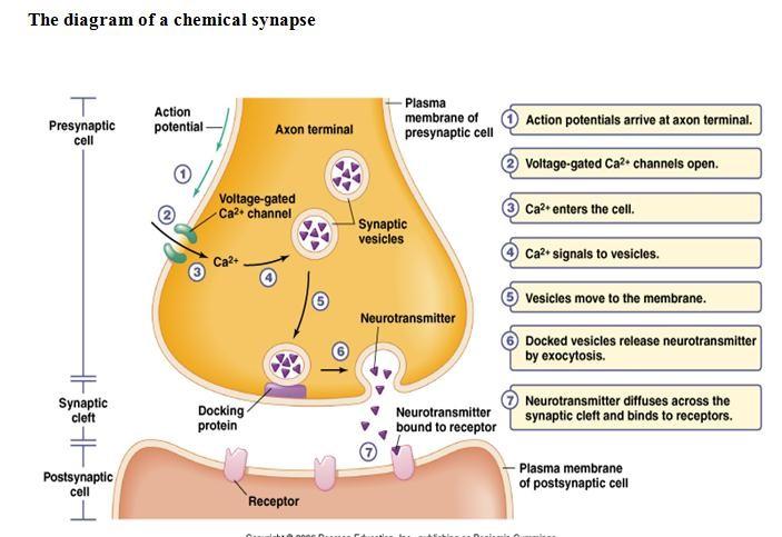
The diagram above shows various events takes place in a chemical synapse which uses acetylcholine as a neurotransmitter.
COORDINATION
Acetylcholine is synthesized within the pre synaptic knob and stored in special organelles called synaptic vesicles (some neurotransmitters neither decrease nor increase the tendency of a postsynaptic cell to fire an action potential instead they work like hormones; e.g. noradrenaline released by axons of the sympathetic nervous system has an effect to on cells similar to that of the hormone adrenaline.
When an action potential reaches the presynaptic membrane, it depolarizes the membrane, that is; it makes the membrane less negative than at rest.
This depolarization triggers the opening of calcium ions channels in the pre synaptic membrane.
COORDINATION
The calcium ions diffuse into the presynaptic knob; causing the pre synaptic vesicles containing acetylcholine to migrate and fuse with the presynaptic membrane.
The acetylcholine is released into the synaptic cleft and diffuses across the synapse.
The synapse then it binds to specific protein receptor molecules on the post synaptic membrane a process known as receptor activities.
Receptor activation causes sodium ion channels to open making the membrane more permeable and produce a graded potential, if enough acetylcholine is released the graded potential may become large enough an action central.
COORDINATION
If an excitatory synapse receives a continuous stream of action potential at high frequency eventually transmission across synapse stops.
This is because the neurotransmitter cannot be resynthesised fast enough and it runs out.
The synapse becomes fatigued (i.e. adapted).
Acetylcholine is then broken down by acetylcholinesterace to its constituents groups acetic acid (or ethanoic acid) and choline groups; which diffuses back into the pre synaptic membrane and used to resynthesize the neurotransmitter under the influence of ATP from the mitochondria concentrated here and refilled in vesicles for future transmission.
Inhibitory synapse.
These release neurotransmitter that makes the postsynaptic membrane less excitable and less likely to transmit an impulse.
E.g. In mammals, they occur in nerve pathways which central rapid eye movements, they are also common in the heart.
COORDINATION
COORDINATION
Diagram above: An electrical synapse in which the nerve impulse is transmitted through the protein pores that line the cytoplasm of the two cells.
Qn; 1. Write short notes on a synapse
SENSORY ORGANS
Receptor cells
These are specialized cells in the body forming the body’s principal means of gaining information about the environment.
Characteristics of receptor cells
They may be either proportions of nerve cells e.g. many sensory endings in the skin OR they may be specialized intimate contact with nerve cells e.g. the cells in the tongue.
COORDINATION
Each type of receptor is responsive to a particular kind of stimulus, stretching, pressure, light.
Most receptors will not respond stimuli other than those for which they are specialized.
Each type of receptor functions as a transducer; converting the energy that constitutes the particular stimulus to which it is attained into the electrochemical energy to the nerve impulse.
Each type of receptor sends impulse to a particular part of the brain.
Mechanism of receptor function.
Qn; By what process do the environment phenomena that constitute stimuli causes receptors to initiate impulses?
COORDINATION
In the previous discussion about the post synaptic depolarization, we saw the excitatory transmitter substance at synapse induce impulse in the post synaptic neuron by reducing the polarization of the membrane of that to a critical level i.e. by inducing an EPSP that reaches threshold potential for impulse generation.
A similar process is involved in the case of sensory receptors; the stimulus causes sufficient depolarization of the membrane of the receptor cells causes it to initiate an impulse.
COORDINATION
The stretching of the muscle spindle produces a local depolarization of a receptor cell.
This depolarization is called generator potential. When the generator potential reaches the threshold level it triggers an action potential in the nerve fibre.
COORDINATION
An increase in intensity of the stimulus causes the proportional rise in generator potential.
This rise in turn and the rate at which it occurs determine the frequency of the triggered impulses.
Thus the output from the receptors conveys a measure of the strength of the stimulus.
Qn; How does the stimulus produce the generator potential?
The membrane becomes more permeable to Na+ ions which thus flow inward.
COORDINATION
Qn; How does the stimulus increase the permeability of the membrane to Na+ ions?
Since different receptors are stimulated by different stimuli the mechanism may as well vary.
e.g. i) In the case of vision, light energy is known to cause chemical change in receptor pigments and these changes are presumed to initiate chemical reactions that produce some sort of transmitter substance may depolarize the membrane of the next cell in the pathway.
COORDINATION
ii) In the case of stretch receptors and pressure receptors mechanical distortion of the membrane is thought to cause a permeability change directly, either by opening channels or through the membrane that allows Na+ to leak inward, or by increasing the size of the channels (for electrical synapses) otherwise too small for the passage of Na+ ions. NB;
The size of the generator potential is directly proportional to the size of the stimulus.
COORDINATION
The direct relationship between strength of stimulus and magnitude of generator potential could be explained by assuming that stronger stimuli distort a greater area of membrane; there are openings, more channels and allowing more ions to pass through the membrane.
THE MAJOR SENSES
In biology, human being have five senses; touch, taste, smell, vision and hearing.
TYPES OF RECEPTORS
Receptors are cells that receive information from the environment and send impulses via conductors to the central nervous system. Receptors may be categorized on the basis of;-
COORDINATION
lower-roman;margin-left: -0.7500000000000036pt;text-decoration: none;vertical-align: baseline”>
Types and function of the stimuli they respond to
Mechanoreceptors-detect movement, pressure or tension.
Photoreceptors-detect variation in light
Chemoreceptors-detect chemicals
COORDINATION
Thermoreceptors-respond to both internal and external heat and cold Pain receptors-respond to tissue damage.
lower-roman;margin-left: -0.7500000000000036pt;text-decoration: none;vertical-align: baseline”>
Complexity of receptor structure
Primary receptors
Secondary receptors Sense organs
lower-roman;margin-left: -0.7500000000000036pt;text-decoration: none;vertical-align: baseline”>
Source of stimulus.
COORDINATION
Exteroreceptors-respond to stimulus outside the body
Interoreceptors- respond to stimuli inside the body
Proprioreceptors- specifically sensitive to the relative positions of the skeleton and degree of muscle contraction.
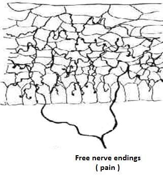
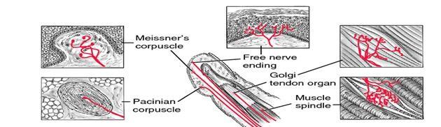
SENSORY RECEPTORS OF THE SKIN
There are numerous types of sensory receptors in the skin which shows some receptors in place.
These receptors are concerned with at least five different senses namely touch, pressure, heat, cold and pain.
Some of the skin receptors particularly those concerned with pain are simply the unmyelinated terminal braches of neurons.
COORDINATION
COORDINATION
Others are nets of nerve fibres surrounding the bases of hairs. These are particularly important in the sense of touch.
These are stimulated by the slightest displacement of the tiny hairs present on most parts of the body.
Other skin receptors are more complex consisting of nerve endings surrounded by a capsule of a specialized connective tissue cells.

COORDINATION
NB;
The relative abundance of the various types of the receptors differs greatly e.g. pain receptors are nearly 27 times more abundant than cold receptors and cold receptors are nearly 10 times more abundant than heat receptors.
The receptors are not evenly distributed over the entire body e.g. touch receptors are much more numerous the finger tips than in the skin of the back.
THE PROPRIOCEPTIVE AND VISCERAL SENSES.
These are widely distributed over the internal body and function primarily in receiving information about the condition of the body itself.
COORDINATION
Though the senses receptors mediate are not included in traditional classification of the five, they are in immense importance in the life of an organism.
Examples are the stretch receptors (proprioreceptors) in the muscles and tendons, which are involved in the knee jerk reflex.
They are sensitive to the changing tension of muscles and tendons and send impulses to the central nervous system in forming it of the position and movements of the various parts of the body.
COORDINATION
The terminal branches of sensory nerve fibre are intimately associated with several specialized muscle fibre that form an apparatus called neuromuscular spindle.
Other dispersed receptors include those of visceral senses located in the internal organs e.g.
COORDINATION
receptor in the carotid artery sensitive to carbon dioxide concentration in the blood and to blood pressure.
The firing of such visceral receptors seldom results in sensations (i.e. we are not aware of their action) the responses to the stimulation of visceral receptors produces conscious sense such as thirst, hunger and nausea.
THE SENSE OF TASTE AND SMELL
The receptors of taste and smell are chemoreceptor i.e. they are sensitive to solutions of certain types of chemicals, which can find to them by weak bonds.
The two sensations are much alike and when we speak about a taste sensation we are not referring to a compound sensation produced by stimulation of both taste and smell receptors.
COORDINATION
One reason why we cannot “taste” food well with a cold is that with nasal passages inflamed and coated mucus, the smell receptors are essentially non functional.
In other words; much of what we call taste is really smell.
Consequently some vapours entering our nostrils pass across the smell receptors and down into the mouth where they stimulate taste receptors.
COORDINATION
In each case taste and smell, chemicals must go into solution in the film of liquid coating the membranes of the receptor cells before they can be detected.
The major functional difference between the two kinds of receptors is that taste receptors are specialized for detection of chemicals present in quantity in the mouth itself while smell receptors are more specialized for detecting vapours coming to the organisms from distant source.
They are much more sensitive that taste receptors as much 3000 times more in some instances.
COORDINATION
One reason why hot foods often have more “taste” than cold food is that they vaporize more.
The vapours passing from mouth upward into the nasal passage and these stimulate smell receptors.
TASTE.
The receptor cells for taste are located in taste buds or the upper surface of the tongue and to a lesser extent on the surface of the pharynx and larynx.
The receptor cells themselves are not neurons but specialized cells with microvillus on their outer ends.
COORDINATION
The end of nerve fibres lie very close to these receptor cells, and when a receptor cell is stimulated, it generates impulses in the fibres.
The picture above shows the structure of the tastebud
(Each test bud contains specialized receptor cells bearing sensory microvilli that are exposed in pits on the tongue surface.
The ends of sensory neurons (coloured) are closely associated with this receptor cells).
SMELL
The receptor cells for the sense of smell (olfaction) in humans are located in two clefts in the upper parts of the nasal passages.
COORDINATION
Unlike the receptor cells of taste, the olfactory receptors re true neurons.
The cell bodies to the surface of epithelium where they bear a cluster of modified cilia, which function as receptor sites.
FUNCTIONAL PROPERTIES OF RECEPTORS.
1. ADAPTATIONS.
COORDINATION
Adaptation is the decline in the frequency of impulses when a strong or constant stimulus is perceived by a sensory receptor cell e.g. on entering a room you may immediately notice a clock ticking but after a while you become unaware of its presence.
The rate and extent of adaptation in a receptor cell is related to its function and there are types; rapidly and slowly adapting receptors.
Slowly adapting receptors (ionic receptors) register constant stimulus with a slowly decaying frequency of impulses.
COORDINATION
The adaptation is thought to be due to decrease in the permeability of the receptor membrane to ions due to substance stimulation.
This progressively reduces the size and the duration of the generation potential and when this falls below threshold level, the sensory neuron ceases to fire the impulse.
Advantage of adaptations
It provides animals with precise information about change in the environment.
COORDINATION
At other times the cells do not send signals thus preventing over loading of the central nervous system with irrelevant and unmanageable information.
This ensures efficiency and economy of the nervous system.
2. RAPIDLY ADAPTING RECEPTOR (PHASIC RECEPTOR)
Respond to changes in stimulus level by producing a high frequency of impulses at the movements when the stimulus switched “on” or “off”
COORDINATION
E.g. Pacinian corpuscle and other receptors concerned with touch and the detection of sudden changes acts in this way.
CONVERGENCE
In sensory receptors, several receptor cells will often synapse with a single receptor neuron as shown in a figure above.
This means that while the generator potential from an individual receptor cell may be insufficient to set up an action potential across the synapse.
COORDINATION
The generator potential from receptor cells may add together or summate and trigger an action potential.
This is known as convergence and is a useful adaptation for increasing the sensitivity of a sensory system to low level stimuli.
THE SENSE ORGAN.
Single sensory receptor cells are very useful and can carry vital information.
But groups of receptor cells specialized for picking up a particular stimulus can be even more useful.
Relatively early in the development of the animal groups’ collection of receptors evolved together to form specialized regions which are called sense organs.
Throughout the animal kingdom, the most common sense organs are those which respond to light and sound or vibrations.
We shall consider in some detail the human eye and ear, with reference to some of the alternative structures which are found in other groups.
THE HUMAN EYE; THE EYE AT WORK.
a) The role of iris
Iris is a circular sheet of muscles dividing the eye into two chambers.
The pigment it contains gives the eye its colour. The reflex contraction and relaxation of the muscles of the iris control the amount of light entering the eye.
When we look at something our eyeballs are moved in their sockets by muscles so that the pupil at the centre of the iris is pointing at the object of our interest.
Light from the object enters the eye through the pupil and the amount of light entering is controlled by the size of the opening.
This in turn is controlled by the Iris muscles.
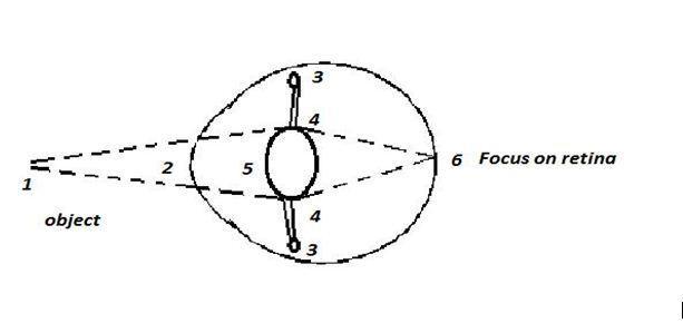
ACCOMMODATION
Accommodation is the reflex mechanism by which light rays from an object are brought to focus on the retina.
It involves two processes which are,
REFLEX ADJUSTMENT OF PUPIL SIZE
1. In bright light
The iris reduces the size of pupil by contracting its circular muscles and relaxing its radial muscles to prevent damage to the light sensitive cells by strong light.
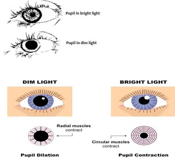
2. In dim light /poor light intensity
The circular muscles relax and the radial muscle contract opening the pupil aperture as wide as possible to the maximum amount of light to ensure the best possible vision.
3.

CHANGING THE SHAPE OF THE LENS.
The ciliary muscles are arranged circularly around the ciliary body, the effects of their contractions and relaxations are relayed to the lens by the suspensory ligaments.
The lens itself is elastic and its unstretched shape is relatively short and fat.
When ciliary muscles relax, the gap around the lens gets longer, increasing the tension in suspensory ligaments.
These in turn pull on the lens making it long and thin.
Its ability to bend light is now minimum and it is said to be unaccommodated.
When ciliary muscles contract they reduce the gap around the lens.
This reduces the tension in the suspensory ligaments allowing the lens to become short and fat.
In this state it is fully accommodated and its ability to bend the light is maximum.
REFRACTION OF LIGHT RAYS
Light rays from a distant object are parallel when they strike the eye.
Light rays from new object are diverging when they strike the eye.
Both cases light rays must be refracted or bent to focus on the retina and refraction must greater for light from near objects.
Refraction occurs when light passes from one medium into another with a different refractive index, and this occurs at the air to the cornea at the surface of the lens.
The degree of refraction at the cornea surface depends on the angle at which light strikes the cornea; also depend upon the distance of object from the cornea.
Most of the refraction occurs in the cornea and consequently the function of the lens is to produce the final refraction that brings light to sharp focus on the retina.
The light interring the eye is refracted by its passage through the conjunctiva, cornea, aqueous humour and vitreous humour in exactly the same way regardless of whether it is from a near or a distant object.
But by changing the shape of the lens the degree of bending of the light can be altered.
Light from distant objects needs relative little bending to bring it into focus on the retina and so the lens has to be thin.
(Because they are almost parallel and not diverging like for near object).
1. Light from a distant object

Parallel light rays reach the eye
Cornea refract (bends) light rays
Circular ciliary muscles relaxes
Suspensory ligament taut
Lens pulled out thin
Light focused on the retina.
1. Light from near object
Diverging light rays reach the eye
Cornea refracts (bends) light rays
Circular ciliary muscles contracted
Suspensory ligament slack
Elastic lens more convex
Light focused on retina
To bring light from near objects into the focus on the retina more refraction is needed and so the lens has to be short and fat.
This ability to focus light from objects of various distances is known as accommodation.
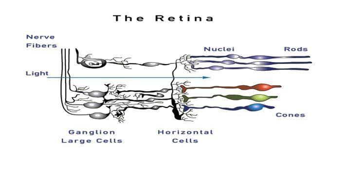
THE ROLE OF RETINA.
Retina is a layer of light sensitive cells (i.e. rods and cones and the neurones leading from these photoreceptors to the optic nerve.
The light from the objects is focused into the retina. The retina must then perceive that light and inform the brain of its presence.
In order to do this the retina contains about a hundred million light sensitive (photoreceptors) along with the neurones with which they synapse.
There are two main types of photoreceptors in the retina known as the rods and the cones; shown in the figure below.


THE STRUCTURE OF THE RETINA
The retina is composed of three layers of cells each containing a characteristic type of cell; these are:
Photoreceptor layer (outermost layer) containing photosensitive cells; the rods and cones partially embedded the pigmented epithelial cells of the choroid.
Intermediate layer containing bipolar neurons with synapse connecting the photoreceptor layer to the cell of the third layer.
-cells called horizontal and amacrine cells found in this layer enable lateral inhibition to occur.
Note;
Those three layers of the retina are arranged anatomically in reverse order from what it might be expected; the receptor cells are in the back of the retina; and light must pass through the ganglion cells and bipolar – cell layers to reach them.
The reason for this somewhat unexpected arrangement is the origin of the retinal cells in the embryo and the way in which the eye is formed during the embryonic development.
To add to this confusion; the optic nerve carrying the visual information cross over on their way to the visual cortex in the brain so that the information seen with the right eye is taken to the left side of the brain for processing.
Qn:
Explain the occurrence of blind spot in the retina.
Explain the mechanism of controlling the amount of light entering the eye.
Answer plan
In bright light
More photoreceptor cells in the retina are stimulated by increase in light intensity.
Greater number of impulses along neurons to the brain.
Brain sends nerve impulses along parasympathetic nervous system to the iris diaphragm In the iris circular muscles contract and the radial muscles relax.
Pupil constricts reducing its size of aperture. Less light enters the eye.
In dim light
Few photoreceptor cells are stimulated due to decrease in light intensity.
Fewer impulses pass along sensory neurons to the brain which sends impulses along the sympathetic nervous system.
In the iris diaphragm, circular muscles relax and radial muscles contract
Pupil relax (dilates)
1. More light enters eye.

A diagrammatic section through the retina of the eye showing the ultra structure of a rod and a cone.
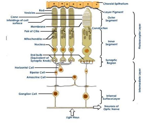
Outer segments
Outer segment is the light sensitive region where the light acts as a stimulus to the production of generator potential
They contain flattened membranous vesicles filled with photosensitive pigments.
Constriction:
Constriction is a very narrow region between the outer and inner segment.
Inner segment
This is packed with mitochondria which produce energy for various processes and ribosome’s which synthesize proteins for the vesicles and visual pigments.
Synaptic region.
The cells from synapses with the bipolar cells and several rods synapse with one bipolar cell join to give increased sensitivity to light (convergence phenomenon).
Once connected with one bipolar cell giving great visual activity.
NB:
Visual acuity: is the ability of the eye to resolve two or more stimuli spatially separated.
The cells of the human retina
The human retina has the following cells.
The receptor cells (rods and cones) making the outer layer.
The bipolar cells which synapse at their tips with receptor cells these make up the in middle layer of retina.
Ganglion cells: Which synapse at their tip with the bipolar cells; make the third layer
-Their axons form the optic nerve, which run from the eyes to the brain.
NB;
Processing the information can occur within the retina because of the several bipolar cells synapses with a single ganglion cells
Besides convergence of information; there is lateral transfer of information from pathway to pathway via horizontal cells (each of which receives synapses from many receptor cells and synapses on many bipolar cells on the other horizontal cells) and via amacrine cells.
(which both receive synapses from and synapse on bipolar cells, and also synapse on many ganglion cells)
Roles of horizontal cells and amacrine cells.
1. Horizontal cells
Synapse with several bipolar neurons; this increases visual acuity and sensitivity of the vision.
By exerting lateral inhibition.
If they receive stimuli from two rods of equal intensity they cancel out (inhibit the stimuli).
They therefore enhance contrast between areas that are strongly stimulated and there that are weakly stimulated.
This makes features such as edge of objects stand cut more clearly.
2. Amacrine cell (are stimulated by bipolar neurons and synapses with ganglion cells.)
They transmit information about changes in the level of illumination.
And hence comment on the mode of action of the two nervous system based on the type of neurotransmitter substance they produce.
The rods and cones synapse in the retina with short sensory neurons (bipolar neurons) which themselves synapse in the retina with longer neurons (ganglion cells) whose axons bundled together as the optic nerve, run to the visual centers of the visual centers of the brain.
o The presence of several sets of synapses within the retina enables the eye to modify extensively the information transmitted from the receptor cells to the b
THE LIGHT SENSITIVITY OF RODS AND CONES.
Both rods and cones contain light sensitive pigments
In rods the pigment, which is built into the membrane of the flattened vesicles in the outer segment as called rhodopsin
Rhodopsin is made up of a protein scotopsin (opsin) rended with a light absorbing prosthetic group called retinene retinal, which is a derivative of vitamin A.
When a molecule of rhodopsin is struck by a photon of light, the retinal is converted into a slightly different isomer i.e.
When converted to Trans- isomer; rhodopsin then breaks up into opsin and retinene.
The breaking of rhodopsin sets up a generator potential in the rod and if this is large enough or if several rods are stimulated simultaneously, an action potential is set up in the receptor neuron.
Once bleaching/ breaking of the rhodopsin have occurred, the rod cannot be stimulated again until rhodopsin is resynthesized.
It takes energy from ATP produced by the many mitochondria in the inner segment to convert retinene back to the cis – isomer and rejoin and opsin.
QUESTIONS
Qn. 1
Explain the structure of a rod and cone cells. Briefly show how each is adapted to its function.
Name the cellular components of retina and state the role of each type of a cell.
Name the various layers making up the retina.
Give evidence of information processing in retina before it is interpreted in the grain. Explain briefly.
Explain the concept of visual acuity and show the reality that cones have high visual acuity than rods.
Qn. 2
a)State any two special anatomical exceptions noted as far as the retina and associated neurons are concerned.
b) Explain what happens in iris and retina when one
Use illustrative diagrams where possible in supporting your answer and explain the biological terms (if any) that applied to any of your answers above.
Qn. 3
a) Explain the process of accommodation based on
Qn. 4
lower-alpha;margin-left: -0.75pt;text-decoration: none;vertical-align: baseline”>
lower-alpha;margin-left: -0.75pt;text-decoration: none;vertical-align: baseline”>
1. Parasympathetic nervous system 2. Sympathetic nervous system.
MECHANISM OF PHOTORECEPTION
Rods contain the photosensitive pigments called Rhodopen or vival putple.
Rhodopsin is made by combination of a protein called satopsin with a small light absorbing molecule called retinene (retired) which is a derivative of vitamin A.
In the presence of lights rhodopsin decomposes into retinene and scetopsin a process known as bleaching.
Rhodopsin is formed in the absence of further stimulation of light a process known as Dark adaptation.
The retinal exist into two isomers.
Bleaching leads to the creation of a generator potential in the rod cell which is sufficiently large, generate on action potential along the neutrons leading from the cell to the brain.
For the daylight cones are used.
COORDINATION
They contain photosensitive pigments called Iodepsin.
PHYSIOLOGY OF SEEING:
The light rays from an object reach the eye and pass through the transparent conjunctive, cornea, aqueous humor and crystalline line.
The cornea bends the light rays and the line causes more bending there refracted light rays passes through the vibreake humour and finally came to focus at a point in the reline.
COORDINATION
The point at which the image focus is called fovea or yellow spot and the image formed is Real, smaller than object and invested.
On the fovea, the light impulse are converted into electrochemical impulses and are sent to the visual area of the brain through the .. nerve.
In the brain an Interpretation of the size, nature, distance and uprightness of the object is made.
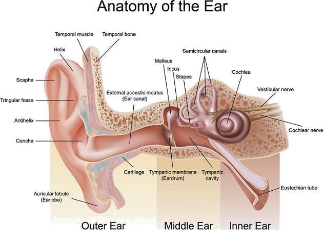
THE MAMMALIAN EAR:
The ear is a sense organ containing mechanoreceptors sensitive to body displacement and sound.
COORDINATION
Movement and portion of the head relative to gravity are detected by the vestibular apparatus composed semicircular canals, succulus and utricle.
All other structures the ear are involved in receiving amplifying and transdusing.
Energy into electrical impulse and promotion of the sensation of the inner ear is principally heating and balancing part of the ear.
COORDINATION
STRUCTURE OF THE MEMBRANEOUS LABYRINTH
Pointed out earlier, the membranous labyrinth is involved in hearing and balance.
It is found in the inner ear.
COORDINATION
It is structurally compound of three semicircular canals that lie, at right angles to one another.
The canals arise from a swollen utricules, Below is high coiled that is involved in hearing. T
he succulus and a connection between the ampula is known as ductus utriaili.
COCHLEA AND HEARING:
Cochlea is spiral sub deviled into three layers vertibular canal and tympanic canal contains perilymph and median canal which contain endolymph.
COORDINATION
The basilar membrane separates the median and tympanic canals and supports sensory hair cell that can be brought into contact with the tecterial membrane above.
This unit consists basilar membrane, sensory cells and tectorial membrane is called ….organ of coit and is the region where transduction of sound … into electrical impulse occurs
COORDINATION

COORDINATION
Organ of Corti.
MECHANISM OF HEARING:
Sound waves are directed toward the inner ear through the External auditory meatices where they cause the tympanic membrane to vibrate.
COORDINATION
In the middle ear the vibration of the tympanic membrane are across the oval window by movement of the three ear asides, the mallcus, incurs and stapes.
The vibration are than transmitted into the innear ear where they cause perilymph of the vestibular canal to vibrate and these are transmitted via Reissner’s membrane to the endolymph in the median canal. From there they are transferred to the basilar membrane and the perilymph in the tympanic canal, and are finally dispirited, into the air of the middle ear as vibration of the round window.
COORDINATION
Vibration of basilar membrane pushes the sensory hair against the tectorial membrane and forces the two membrane to slide part each other.
The distortion produced in the sensory hair cells due to the shearing forces causes a depolarization of the sensory cells, the production of generator potential, and initiation of action potentials in the axons of the auditory nerve.
COORDINATION
The latter transfer the impulse to Auditory part of the brain where an interpretation of the pitch note, intensity and quality of the sound is mode.
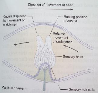
THE MAMMALIAN EAR AND BALANCE:
Several parts of the body are involved in maintaining balance at … and during movements. The parts that are involved include eyes, …. Receptors.
In joints and muscles etc. however vital information, ….. to position and movements of the head is provided by the vestibular apparatus of the ear, the utricle, sucaule and semicircular canals.
COORDINATION
The basic sensory receptor in these structure consist of the hair cells attached to dense structures supported in the Endolymph, the region of the walls of utricle and saccule, the maculae contains granules called atoconia in association to receptor cells.
The atoconia responds to gravitational pull and mainly detect the direction of movement of the head with respect to gravity.
COORDINATION
The utricle respond to vertical movement of the head e.g. when the body is upside down.
The saccule responds to lateral movement of the head.
The semicircular canals responds to rotational movements of the head and they contains cupules that works in the same way of maculae.
Linear acceleration is detected by both maculae and ampullae.
COORDINATION

COORDINATION
Ampulla of semi-circular canal Membranous labyrinth of human
N.B: Adaptation of organ of corti
lower-roman;margin-left: -0.75pt;padding-left: 3pt;text-decoration: none;vertical-align: baseline”>
lower-roman;margin-left: -0.75pt;padding-left: 3pt;text-decoration: none;vertical-align: baseline”>
COORDINATION
lower-roman;margin-left: -0.75pt;padding-left: 3pt;text-decoration: none;vertical-align: baseline”>
lower-roman;margin-left: -0.75pt;padding-left: 3pt;text-decoration: none;vertical-align: baseline”>
HORMONAL CO-ORDINATION:
COORDINATION
In mammals and other higher animals there are two co-coordinating system those include nervels system and hormonal system.
Defn: A hormone is a chemical substance which is produced at one part of the body and exerting it’s effect at another part of the body away from it’s centre of production.
The organ/part that receives effects of hormones are called target organe or target parts.
The hormone is secreted directly into the blood stream.
Such ductless gland which secrete hormone are called endocrine glande which constitute the endocrine system.
Mechanism of Hormone Action:
COORDINATION
The mechanism of controlling the release of Hormones by the glands is as follows:-
Presence of specific metabolite in the blood, e.g. Presence of Excess Glucose in the blood, causes the release of Insulin
Presence of another hormone in the blood e.g. Many of hormones released by the anterior pituitary cases the release of other hormones.
Stimulation by neurons from autonomic nervous system e.g. adrenaline and noradreline are released by the Adrenal gland following the arrival of impulses in the condition of anxiety, stress and danger
COORDINATION
FEEDBACK MECHANISM OF HORMONAL CO-ORNATION:
Feedback mechanism is a self regulating mechanism in the body which tends to restore the physiological equilibrium or stability of the body or increase in the instability of the body.
TYPES OF FEEDBACK:
(a (a) Positive Feedback:
This is a type of Feedback that tends to increase the effect of the disturbance.
Positive feedback responses are rare in biological systems because they increase instability of the body.
(b) Negative Feedback:
COORDINATION
This brings the body back to its normal physiological states thus the negative feedback mechanism, are important in the biological systems.
– Examples of biological negative feedback mechanisms include the control of heartbeat, blood pressure, body temperature and hormone levels i.e. secretion of insulin.
N.B:
TRF – Thyroid releasing factor
TSH – Thyroid stimulating hormone
COORDINATION
Note: Examples of positive feedback:
During labour when hormone oxytocin stimulate muscular contraction of the uterus which in turn stimulate the release of more oxytocin.
Interaction between Hormonal and Nervous Systems:
The two co-ordinating systems i.e. hormonal and nervous system at one point depend on one another.
For example the release of hormone sometimes depend on the response reaching the Gland through the nerve cell.
For example adrenalin and neradreline are released by the adrenal gland following the arrival of impulse in conditions of danger, anxienty and stress.
COORDINATION
Thus hormonal and nervous system depends one another when co-ordinating the body system.
NERVOUS AND HORMONAL CONTROL OF SECRETION:
In mammals the production of digestive secretion is under both nervous and hormonal control.
The following are number of juices secreted and the actions of hormones and nervous towards secretions.
COORDINATION
SALIVA:
As above explained sight, smell and thought can lead to secretion of saliva.
Also presence of food in the tongue stimulate further recreation of saliva.
Note: There are three important hormones secreted in the stomach and small intestinal region called:-
lower-roman;padding-left: 44.3pt;text-decoration: none;vertical-align: baseline”>
Gastrin
COORDINATION
lower-roman;padding-left: 44.3pt;text-decoration: none;vertical-align: baseline”>
lower-roman;padding-left: 44.3pt;text-decoration: none;vertical-align: baseline”>
GASTRIN:
Presence of food in the stomach stimulates the walls of stomach to pralls a hormone called “ gastrin” which passes into the blood stream.
COORDINATION
Gastrin stimulate the production of gastric juice from gastric gland for up to 4Ø occurs
Note: Digestion of fats takes longer and requires less acidic conditions ….. sence of fats in the stomach initials the production of hormone called terogesterone which inhibits any further secretion of the acid by oxyntic of the stomach.
SECRETIN:
This hormone is produced by the small intestinal wall cells and passes the blood stream and meets the three target liver, stomach and increase.
COORDINATION
IN THE STOMACH: It inhibits the secretions of gastric juice by inhibiting the secretion of gastrin
IN THE LIVER: It stimulates the bile to produce salts like NaHCO2 used in neutralization of ocidic style chime.
IN PANCREASE: It stimulates the pancreas to produce alkaline salts e.g. NaHCO3
CHOLE CYSTOKININ/PANCRE: OZYMIN
This hormone also it recreated by small intestinal wall cells and …. into three targets; liver, stomach and pan crease.
COORDINATION
* IN THE STOMACH; Inhibits stomach emptying
*IN THE PANCREASE: It stimulates the secretion of pancreatic juice to the duodenum.
*IN THE LIVER: It stimulate the contraction of gall bladder walls hence secretion of bile through bile through bile duct.
EPITHELIAL AND GLANDULAR TISSUES IN RELATION TO THEIR DIGESTIVE ROLES:
Epithelial tissue is a type of tissue which covers the external parts of the body as well as internal parts, such as lining of atemosmestory canal.
Epithelium performs various functions such as diffusion in transportation and secretion of substances as well as protection of the body parts
Example of Epithelial tissue.
COORDINATION
(a Columnar Epithelial tissue:
This consists of elenejated calls which are quite narrow, thus providing more cytoplasm per unit area of epithelium.
Each all possesses a nucleus situated at its basal end. Also it certain goblet cells:
There is a striated border or brush border of microvillus at the force surface end of each cell.
Location: stomach small intestine, kidney ducts and thyroid gland.
Functions:
COORDINATION
lower-roman;padding-left: 39pt;text-decoration: none;vertical-align: baseline”>
Mucus secreted by goblet cells in the epithelium of stomach protects the stomach lining from the acidic contents of the stomach and from digestion by enzymes.
lower-roman;padding-left: 39pt;text-decoration: none;vertical-align: baseline”>
Mucus secreted by goblet cells in the epithelium of small intestine protect it from self digestion and lubricates the passage of food
lower-roman;padding-left: 39pt;text-decoration: none;vertical-align: baseline”>
Comparison:
Comparison of Hormonal and Nervous system:
COORDINATION
THE ENDOCRINE GLANDS
There are ductless glands that secrete Hormones directly into the blood stream. Such glands include.
THE PITUITARY GLAND:
COORDINATION
Pituitary gland, also known as master gland is divided into two ….
Pasterior pituitary:- This does not synthesize any hormone but … and release two hormones, Ant diuretic hormone (ADH) and ………which are produced by neurosecretory cell bodies lying in hypothalamus and pass down the nerve fibres.
lower-roman;margin-left: 36.00000000000001pt;padding-left: 41.65pt;text-decoration: none;vertical-align: baseline”>
Is released in response to a in the water content of blood plasma and leads to an increase ..meability of water of the distal and collecting tubules of the … in the kidney so that water is retained in the blood plasma, reduced volume of urine is excreted.
lower-roman;margin-left: 36.00000000000001pt;padding-left: 41.65pt;text-decoration: none;vertical-align: baseline”>
OXYTOCIN: – Cause contraction of uterus during birth
– Stimulate ejection of milk from the nipple
Anterior pituitary:- This is connected to the hypothalamus by blood vessels which form potal system.
COORDINATION
Pituitary gland produce and store hormones known as trophic hormones.
A trophic hormone is one which stimulate other endocrine gland to release their hormone.
Anterior pituitary is regarded as a master gland because it controls secretion of hormone from other glands
Interior of hormone secreted by anterior pituitary gland are controlled by hypothalamus hormones such as:-
lower-roman;margin-left: 36pt;padding-left: 18pt;text-decoration: none;vertical-align: baseline”>
lower-roman;margin-left: 36pt;padding-left: 18pt;text-decoration: none;vertical-align: baseline”>
lower-roman;margin-left: 36pt;padding-left: 18pt;text-decoration: none;vertical-align: baseline”>
COORDINATION
lower-roman;margin-left: 36pt;padding-left: 18pt;text-decoration: none;vertical-align: baseline”>
lower-roman;margin-left: 36pt;padding-left: 18pt;text-decoration: none;vertical-align: baseline”>
lower-roman;margin-left: 36pt;padding-left: 18pt;text-decoration: none;vertical-align: baseline”>
COORDINATION
Prolactin releasing factor – This stimulate release of prolactin (vii) Prolactin inhibiting factor – This inhibits the release of prolectin.
HORMONES OF THE ANTERIOR PITUITARY:
The hormones secreted by the anterior pituitary include the following:-
lower-roman;margin-left: 36pt;padding-left: 44.3pt;text-decoration: none;vertical-align: baseline”>
Thyroid stimulating Hormone (TSH)
Roles:- Stimulate the growth of thyroid gland
Stimulate T. gland to release thyroxine hormones
lower-roman;margin-left: 36pt;padding-left: 44.3pt;text-decoration: none;vertical-align: baseline”>
Adrenal corticotrophin Hormones (ACTH)
Roles:- It regulates growth of the adrenal cortex
Stimulates adrenal cortex to release hormones such as adrenaline.
lower-roman;margin-left: 36pt;padding-left: 44.3pt;text-decoration: none;vertical-align: baseline”>
Follicles stimulating Hormone (FSH)
Roles:- Stimulate the development of Gracifian follicles
Initiates sperm formation in the testis
lower-roman;margin-left: 36pt;padding-left: 44.3pt;text-decoration: none;vertical-align: baseline”>
Luterising Hormone (LH)
Roles:- Stimlates ovulation and formation of corpus luterising
Stimulates secretion of testerone from the cells in to…
lower-roman;margin-left: 36pt;padding-left: 44.3pt;text-decoration: none;vertical-align: baseline”>
Prolactin (Luteotrophic hormone) LTH
Roles:- Maintain progesterone production from the corpus luterim
– It induces milk production in pregnant females.
lower-roman;margin-left: 36pt;padding-left: 44.3pt;text-decoration: none;vertical-align: baseline”>
Growth hormone (GH) or somatotrophin Hormone (STH)
Roles:- Promotes growth of the skeleton and muscles
– Controls protein synthesis and general body metabolism
Stimulates the production of glucagon by the X-Cells of islets of langerhane of the pancreases
Abnormality of Pituitary Gland:
lower-roman;padding-left: 18pt;text-decoration: none;vertical-align: baseline”>
Deficiency (hypor secretion) of growth hormones before maturity leads to dwarfism.
lower-roman;padding-left: 18pt;text-decoration: none;vertical-align: baseline”>
Overproduction (hyper secretion) of growth hormones during skeretal development results to Giantism (gigantism)
Excess of Growth hormones at maturity causes excessive growth of certain part of the body such as palm of the hand. This called Acremegally
THE THYROID GLAND:
This gland is found in the neck region in each side if the junction …… the Larynx (veils box) and trachea (wind pipe)
Hormones of the thyroid gland:
Trilodo thyroxine (T3)
Tryroxine (T4)
Calatorin
T3 and T4 are similar structurally and functionally they differ the fact that T3 contains three Iodine atoms while T4 4 Iodine atoms
(i) To control the basal metabolic rate (BMR):- Basal metabolic rate is the rate at which oxygen and food are used to release energy. It is rate at rest. (ii) To promote the breakdown of glucose and fats to provide energy
lower-roman;margin-left: 36pt;padding-left: 18pt;text-decoration: none;vertical-align: baseline”>
Thyroxine and growth hormone 9GH) has joint Ermlattatory effect on protein synthesis, leading to an increase in growth rate, particulary of the skeletal system
lower-roman;margin-left: 36pt;padding-left: 18pt;text-decoration: none;vertical-align: baseline”>
Thyroxine stimulate brain development 73 and 74 work in conjuction with insulin, Adreneline and glucose corticoid
Calatorin
Roles:- Controls calcium metabolism
Abnormalities of the Thyroid gland:
(a) Hypothyroidism (Under activity)
In immature creatures, Its result into a condition called cretin
A cretin has the following features:-
lower-roman;margin-left: 35.999999999999986pt;padding-left: 44.45pt;text-decoration: none;vertical-align: baseline”>
Dwarf
lower-roman;margin-left: 35.999999999999986pt;padding-left: 44.45pt;text-decoration: none;vertical-align: baseline”>
Mentally retardat
lower-roman;margin-left: 35.999999999999986pt;padding-left: 44.45pt;text-decoration: none;vertical-align: baseline”>
Irregular development of bones and musdes
(iv)
(v) Skin become dry the eyes are puffy, hair is brittle and shoulder are sag.
Myoxedema – Results when the thyroid gland become underactive during adult hood. This condition results into:- (i) Swallon facial features
lower-roman;margin-left: 36pt;padding-left: 41.650000000000006pt;text-decoration: none;vertical-align: baseline”>
Tiredness
lower-roman;margin-left: 36pt;padding-left: 41.650000000000006pt;text-decoration: none;vertical-align: baseline”>
Passible mental retardation
lower-roman;margin-left: 36pt;padding-left: 41.650000000000006pt;text-decoration: none;vertical-align: baseline”>
Intolerance to cold, due to law basal metabolic rate (BMR)
GOITRE, Results due to insufficient production of thyroxine hormone, the thyroid gland swells. This swelling in the neck .. caused by insufficient Iodine in the diet which force the thyroid to expand in an effort to produce more thyroxine
Over activity of the thyroid gland (Hyper thyroidism)
Over activity may be due to overproduction of thyroxin from an …. Thyroid gland.
The symptoms are:-
lower-roman;margin-left: 35.999999999999986pt;padding-left: 44.45pt;text-decoration: none;vertical-align: baseline”>
Increase in heart rate
lower-roman;margin-left: 35.999999999999986pt;padding-left: 44.45pt;text-decoration: none;vertical-align: baseline”>
Increase in ventilation rate
lower-roman;margin-left: 35.999999999999986pt;padding-left: 44.45pt;text-decoration: none;vertical-align: baseline”>
Increase in body temperature
COORDINATION
lower-roman;margin-left: 35.999999999999986pt;padding-left: 44.45pt;text-decoration: none;vertical-align: baseline”>
The basal metabolic rate may increase by 50% with associated
lower-roman;margin-left: 35.999999999999986pt;padding-left: 44.45pt;text-decoration: none;vertical-align: baseline”>
Increase in oxygen consumption and hear production
lower-roman;margin-left: 35.999999999999986pt;padding-left: 44.45pt;text-decoration: none;vertical-align: baseline”>
Extreme hyperthyroidism is thyrotoxicosis and is associated with released excitability of cardiac muscle which my lead to heart lure failure.
PARATHYROID GLAND
These are four tiny gland embedded in the thyirod gland. They stimulate a single hormone called parathormone
– it maintain the level of calcium in the blood at a sufficiently required amount. Hence maintaining proper working of the muscle and nerve.
ADRENAL GLAND:
COORDINATION
These are pair of adrenal glands located one just above each kidneys. The outer region is called cortex and inner region is called Medulla.
Adrenal gland secrete several hormones which effect body metabolism.
COORDINATION
Adrenal cortex produce two types of hormones
Mineralocorticoids e.g. Aldosterone
lower-roman;margin-left: 36pt;padding-left: 41.650000000000006pt;text-decoration: none;vertical-align: baseline”>
Controls water and salt content of body stimulating cation pumps in membrane to conserve Na-1 and Cl– and remove K+
COORDINATION
lower-roman;margin-left: 36pt;padding-left: 41.650000000000006pt;text-decoration: none;vertical-align: baseline”>
Prevent excessive Na+ loss in seat, saliva and urine
lower-roman;margin-left: 36pt;padding-left: 41.650000000000006pt;text-decoration: none;vertical-align: baseline”>
Maintain asmotic concentration of body fluids at a steady state.
Glucocorticoids e.g. costisol
COORDINATION
lower-roman;margin-left: 36.000000000000014pt;padding-left: 39.05pt;text-decoration: none;vertical-align: baseline”>
Promote gluconeogenosis, linier glucagen formation and raise blood glucose level
lower-roman;margin-left: 36.000000000000014pt;padding-left: 39.05pt;text-decoration: none;vertical-align: baseline”>
Promote breakdown of plasma, protein and increase availability of amino acids for enzyme synthesis in the liver
COORDINATION
lower-roman;margin-left: 36.000000000000014pt;padding-left: 39.05pt;text-decoration: none;vertical-align: baseline”>
Prevent inflarrunatory and allergic reactions
lower-roman;margin-left: 36.000000000000014pt;padding-left: 39.05pt;text-decoration: none;vertical-align: baseline”>
Decrease antibody production
Adrenal medulla produce two hormones which are adrenaline and noradrenaline
COORDINATION
The roles of adrenaline and noradrenaline are that they .. body for action and therefore sometimes called fight, flight fear hormones as they allow the body is react quickly to urgencies.
THE PANCREASE
The pancrease rest just below the stomach. It 99% exocrine and endocrine gland. Hormone s secreted by the pancrease in a duster called islet of langerhane . the hormones are:-
COORDINATION
Insulin:- Secreted by the B-cells of the islets of langerhane is hormones converts Glucose into Glycogen i.e. It regulate the amount of sugar in the blood.
Glucagon:- Secreted by the X-cel of lolet of langerhane. This increase the blood glucose level by transmitting the liver to convert glycogen into glucose.
DISORDER OF THE PANCREASE
Diabates Mellitus:
COORDINATION
This is caused by insufficient production of insulin from the ß -cells islets of langerhans, glucose accumulates in blood and is deposited in the kidney but the glucose never enters the cells Symptoms:
Sugar in Urine:
Hardening of arteries
High degree of dehydration and thirty
(F) GONADS:
These Includes:
The tests:- Male gonads
COORDINATION
These secretes the sex hormones called testosterone from the interstitial cells also it stimulates the development of sexual secondly features
Eg. Developments of beard and moustache, penlarge genital organs using and broken voice and increase sexual desire.
Desecration of testosterone leads to decrease size and activity of reproductive organ penis erection and volume of ejaculation.
Secondary sexual features are also affected:
The Ovarice – Female genads
secretes hormones like:-
COORDINATION
Oestrogen the development of sexual secondary features of females. The development and enlargement of female reproductive organs soft voice, the growth of pubic hair and in the armpit and start of menstrual cycle.
Progesterone:
Progesterone:- Is secreted by the corpus luteram it inhibits the release of egg and it stimulate thickening of uterus wall.
COORDINATION
LOCATION OF HORMONE IS IMPROVING HUMAN LIFE AND ANIMAL HUSBANDRY: synthetic hormones has been of great use in improving life and animal husbandry.
Example:
Growth hormone have been much used to induce rapid growth animals such as chicken, cow etc.
Some of the reproductive hormones are used to induce increased production of eggs by chicken.
Hormones like prolactin and oxytocine are used to induce caused mil production in cows and goat etc.
In man the synthetic oxytocin is used to induce labour pain the process of birth.
COORDINATION
Synthetic sex hormones such as progesterone hormone are used as contraceptive method. The contraceptive pills contains progesterone
CO-ORDINATION IN PLANTS:
Its pointed out earlier plants are less irritable than animals because they have only one co-ordinating system i.e. Hormonal co-ordination constituting the phytohormones. Their responses are therefore slow and they often involve growth. Growth in turn can result in movement of organ.
The responses that are indicated by plants involves movement and they are due either external or internal stimuli.
COORDINATION
The response that are due to internal stimuli are called spontaneous responses
Example protoplasmic streaming of cell e.g. In Amoeba.
Responses due to external stimuli are called induced responses.
TYPES OF MOVEMENTS WITH RESPECT TO RESPONSES:
The types of movements exhibited by plants and plant like, organism due to response include the following:-
lower-alpha;margin-left: 72pt;padding-left: 7.200000000000003pt;text-decoration: none;vertical-align: baseline”>
COORDINATION
Tactic movement
lower-alpha;margin-left: 72pt;padding-left: 7.200000000000003pt;text-decoration: none;vertical-align: baseline”>
Nastic movement
lower-alpha;margin-left: 72pt;padding-left: 7.200000000000003pt;text-decoration: none;vertical-align: baseline”>
Tropic movement
COORDINATION
TACTIC MOVEMENT:
A taxis is the process whereby an organism move from one place to another in response to external stimuli. Such a movement is referred to as Tactic movement.
Where the whole of an organism shift from one place to another in response to external stimuli. This movements is not confirmed to plants, it is exhibited by animals and plant like euglena.
COORDINATION
If an organism move towards the source of stimuli, it is said to exhibit positive tactic movements, but if it moves away from stimulus it is said to exhibits negative taxis.
TYPES OF TAXES:
(a) Photo taxis – The organism movies in response to the stimulus of light
e.g. cockroach moves away from light (Negative phototaxis or towards light (positive phototaxis) e.g. Euglera.
lower-alpha;margin-left: 18pt;padding-left: 4.900000000000006pt;text-decoration: none;vertical-align: baseline”>
COORDINATION
Chemotaxis – the organism in response to the stimulus of chemical
lower-alpha;margin-left: 18pt;padding-left: 4.900000000000006pt;text-decoration: none;vertical-align: baseline”>
Positive chemotaxis – sperme of messes and ferns swim towards substances released by ovum
– Negative chemotaxis – Mosquito avoid insect repellent
lower-roman;margin-left: -32.5pt;padding-left: 3pt;text-decoration: none;vertical-align: baseline”>
Aerotaxis – The organism moves in response to stimulus of air (oxygen) Example motile aerobic bacteria move towards oxygen.
COORDINATION
lower-roman;margin-left: -32.5pt;padding-left: 3pt;text-decoration: none;vertical-align: baseline”>
Geotaxis – The organism moves in response to the e.g. plarula larvas swim towards sea bed
lower-roman;margin-left: -32.5pt;padding-left: 3pt;text-decoration: none;vertical-align: baseline”>
Rheotaxis – The organism move in the response to the stimulus of resistance. E.g planaria move against water current
lower-roman;margin-left: -32.5pt;padding-left: 3pt;text-decoration: none;vertical-align: baseline”>
Megnetotaxis – The organism moves in response to the stimulus of magnetic field. E.g. certain motile bacteria.
COORDINATION
Significance of Tactic Movements: Enable organisms to escape unfavourable environment
e.g: Euglena moves away from excess light it avoids the effect of light it such movements increase survival chances of an organism.
Enable an organism to move towards the required material e.g. bacteria to move towards oxygen
They bring together into contacts some cells e.g. sperm and ova (chemotoxis) whose fusion leads to the formation of zygote.
NASTIC MOVEMENT:
A nasty is a process where by a fixed part of stationary plant exhibits non-directional movement in response to external stimulus
COORDINATION
(A) TYPES OF NASTIC MOVEMENT:
Nyctinasty:
These are sleep movement of certain parts of plants such … flowers and leaves where they can close and open on …………………
Forms of nyctinasty:
lower-roman;margin-left: 18.000000000000014pt;padding-left: 44.3pt;text-decoration: none;vertical-align: baseline”>
Photonasty – sleep movement due to stimulus of light
lower-roman;margin-left: 18.000000000000014pt;padding-left: 44.3pt;text-decoration: none;vertical-align: baseline”>
COORDINATION
Thermonasty – sleep movement due to stimulus of temperature
lower-roman;margin-left: 18.000000000000014pt;padding-left: 44.3pt;text-decoration: none;vertical-align: baseline”>
Hyponasty – The type of movement in which some flowers
e.g. tulip close at night by the flower side of the petals growing more rapidly
lower-roman;margin-left: 18.000000000000014pt;padding-left: 18.049999999999997pt;text-decoration: none;vertical-align: baseline”>
Epinasty – The type of movements in which opening is caused by more rapid growth of the upper side of the petals
COORDINATION
lower-roman;margin-left: 18.000000000000014pt;padding-left: 18.049999999999997pt;text-decoration: none;vertical-align: baseline”>
Haptonasty – Type of nastic movement in which the stimulus is touch
e.g. closure of the leaves of the Mimasa pudica on being touched Seismonasty – This is the nastic movement in which stimulus is stuck
(B) MECHANISM OF NASTIES:
Consider the leaves of the mimosa plant, these have swelling called palvinus at the base of a petiole or leaflet which posses large parenchyma cells. Rapid tiger pressure changes in these cells result in the pulvina acting as a hinge joint and bring about movement.
COORDINATION
The stimulus is said to be transmitted by a hormone moving through the xylem, electrical changes are associated with its passage but there are no nervous system.
Significance of Nastic movement.
lower-roman;margin-left: 18.000000000000014pt;padding-left: 17.299999999999997pt;text-decoration: none;vertical-align: baseline”>
Defence of the leaves or plant against external enemies such as high temperature etc.
lower-roman;margin-left: 18.000000000000014pt;padding-left: 17.299999999999997pt;text-decoration: none;vertical-align: baseline”>
COORDINATION
Opening of petals which increase the chance for pollination.
TROPISM:
Tropic movements, these are the directional movements exhibited by the fixed part of a stationary plant in response to external stimulus.
TYPES OF TROPIC MOVEMENT:
Phototropism – Response due to light
Emotropism – response due to chemical
Hydrotropism – response due to water
COORDINATION
Thigmotropism/haptotropism – response due to touch
Theotropism – response due to gravity
Significance of Tropism
Positive phototropism of plant sheets enhance exposure of leaves to light is enable photosynthesis to take place
Positive hydrotropism of roots increases the chance for water absorption plant.
Positive geotropism of roots increases the chance for mineral absorption plant and encourage ……… of plant in the soil
COORDINATION
Positive chemotropism e.g. movement of pollen tubes in response to chemical used at micropyle of ovule enable fertilization to occur hence … of seeds and fruits
PHYTOHORMONES:
These are plant hormones which are commonly known as plant with substances or regulators. They can be either stimulating growth stimulants or retarding growth i.e. growth retardants.
TYPE OF PHYTOHORMONES:
Auxin
COORDINATION
Gibberellins
Cytokirin
Absassic acid
Ethylene
The first three are growth stimulants and last two are growth retardants
A: AUXINS:
These are the prime plants growth hormones. They are synthesized the roots and shoot apices as well as in the young leaves.
COORDINATION
The commonly known group of hormones includes. The indole acetic acid (I.A.A)
Role of Auxins
They promote growth of the roots at very low concentration.
They promote cell elongation in region behind apex, hence stein growth.
The promote growth of roots from cuttings.
They promote fruit growth and sometimes induce pethenocarpy
COORDINATION
They promote apical dominance i.e. They inhibit lateral bud growth
They inhibit abscission. Hence prevent the pre-mature falling of leaves and fruits.
COMERCIAL APPLICATION OF AUXINS:
Promoting- They help in natural setting in the absence of pollination (parthenocapy)
Acoine promote development of root in cuttings also weed killers Selective they are used as selective weed killers they kill broad leaved species.
GIBBERELLINS:
COORDINATION
Gibberellins derive their name from the fungus called gibberella Fujikuroi which causes abnormal elongation of the rice condition called foolish speedling. The commonly known gibberellins is the Gibberellic acid (G.A.)
Plants: GA are abundant in young expanding organ where synthesized at young apical leaves, buds, seeds and roots tips of Gibberellins.
COORDINATION
it promote cell enlargement in the presence of auxine it promote cell division in apical menstems and cambium. it promote fruit growth and sometimes paxthenocapy, it break bud and seed dormancy.
COORDINATION
it promote flowering in long day plants and inhibit in short plants.
COMMERCIAL APPLICATION:
Promote fruit setting and are used for growing seedless grayer element of seedless fruits can sometimes occur in fertilization in place and is known as parthenocaspy
Are used in the brewing industry to stimulate -amylase in barley and hence promote malting.
use of anti-gibberellins” result in short (dwarf), study plant keep green leaves and sometimes greater pest and disease resistence
CYTOKININ:
COORDINATION
Are most abundant where rapid cell division are occurring especially goods where there is embryo growth and development.
Role of Cytokinln
Promote Cell division in the presence of auxins promote fruit growth
Promote note lateral but growth
They break seed and bud dormancy in the presence of light and high temperature
It delay leaf senescence.
COORDINATION
Commercial application:
They are used to prolong life of fresh leaf crops such as cabbage
They are used to keep flowers fresh
They are used to break dormancy of some seeds
ABSCISSIC ACID:
These is synthesized in the leaves, stem and seeds. It is a major plant growth inhibitor. It is sometimes referred to as “abscissia hormone”
It is antagonistic to all other classes of plant growth promoters
Role of Abscissa acid (ABA)
Inhibit stem growth especially during physiological stress e.g. in drought and water logging they promote bud dormancy
COORDINATION
Promote abscission
Commercial application of ABA
Can be sprayed on tree crops to regulate fruit drop at the season. This removes the need for picking over a long
Can be sprayed to the plant to increase closure of the stomata during drought.
COORDINATION
ETHYLENE (ETHENE)
These gaseous compound which is transported in solution form in plant
Growth of the stem notably during the period of physiological stress
Promotes root growth
Promotes bud dormancy
Promotes flowering in pineapple
Promotes fruit ripening
Commercial application of ethane Induces flowering in pineapple
stimulates ripening of tomatoes and citrus fruits it is applied to rubber plants to stimulate the floor of latex.